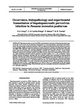
Occurrence, histopathology and experimental transmission of hepatopancreatic parvovirus infection in Penaeus monodon postlarvae
Associated URL
www.int-res.comDate
2003Page views
176Metadata
Show full item recordCited times in Scopus
Share
Abstract
Hepatopancreatic parvovirus (HPV) was detected in samples of Penaeus monodon postlarvae (PL-13, PL-18, PL-19, PL-26) from 2 hatcheries in 2 provinces (Samar and Iloilo) in the Philippines. The percentage of infection was 20 to 100% in postlarvae obtained from the hatchery in Samar in August 2001. Postlarvae from the hatchery in Iloilo, sampled in October and November 2001, had 70 to 99% HPV infection. Wet mounts of squashed hepatopancreatic tissue stained with malachite green (wet-mount technique) and histopathology revealed the presence of large, usually single, basophilic intranuclear inclusion bodies in the distal tubules, which led to displacement of the nucleoli. Light microscopy showed ovoid to spherical inclusion bodies, 5 to 11 µm in diameter. Transmission electron microscopy revealed that the inclusion bodies were composed of electron-dense granular material and virions. The virions appeared roughly spherical and averaged 18 to 22 nm in diameter. An experiment was undertaken to induce HPV infection by feeding P. monodon postlarvae with virus-infected postlarvae. P. monodon postlarvae (PL-16), initially determined as free from HPV, were found HPV-positive 24 h after being fed with infected material. The percentage of infection ranged from 30% at Day 1 post-infection (p.i.) to 100% at Day 7 p.i. determined by the wet-mount technique and by histopathology. This is the first report of a successful horizontal transmission of HPV in P. monodon postlarvae.
Suggested Citation
Catap, E. S., Lavilla-Pitogo, C. R., Maeno, Y., & Traviña, R. D. (2003). Occurrence, histopathology and experimental transmission of hepatopancreatic parvovirus infection in Penaeus monodon postlarvae. Diseases of Aquatic Organisms , 57(1-2), 11-17. https://doi.org/10.3354/dao057011
Subject
Taxonomic term

Collections
- AQD Journal Articles [1215]
Related items
Showing items related by title, author, creator and subject.
-
An overview of the nutrition, feed and feeding techniques of prawn penaeid/shrimps
(Philippine Council for Aquatic and Marine Research and Development, 1989)This paper echoes what transpired during the first International Conference of Penaeid Prawns/Shrimps held in Iloilo City in December 4-7, 1984, particularly on the Nutrition nd Feed Development. Around 25 papers were ... -
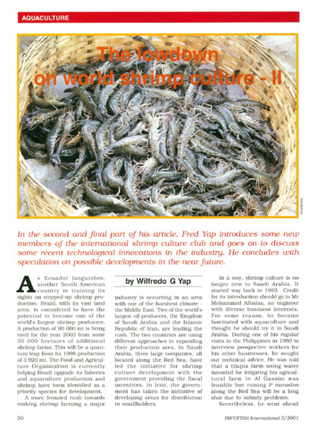
The lowdown on world shrimp culture - II
(INFOFISH, 2001)This paper introduces some new members of the international shrimp culture club and goes on to discuss some recent technological innovations in the industry, particularly the polyculture of tilapia (mainly Oreochromis ... -
Prawn hatchery operations
(Aquaculture Department, Southeast Asian Fisheries Development Center, 1991-02)The manual, an updated version of the 1984 SEAFDEC/AQD manual, presents the underlying principles and step-by-step instructions of prawn larval and post-larval rearing. The techniques described are not only applicable to ...