အရာများအတွက် ရိုးရှင်းသော မှတ်တမ်းများကို ပြပါ။
Nutrition-related histopathology of the intestine of milkfish Chanos chanos fry
Share
| dc.contributor.author | Segner, Helmut | |
| dc.contributor.author | Burkhardt, Patricia | |
| dc.contributor.author | Avila, Enrique M. | |
| dc.contributor.author | Juario, Jesus V. | |
| dc.contributor.author | Storch, Volker | |
| dc.date.accessioned | 2013-01-10T09:35:23Z | |
| dc.date.available | 2013-01-10T09:35:23Z | |
| dc.date.issued | 1987 | |
| dc.identifier.citation | Segner, H., Burkhardt, P., Avila, E. M., Juario, J. V., & Storch, V. (1987). Nutrition-related histopathology of the intestine of milkfish Chanos chanos fry. Diseases of Aquatic Organisms, 2(2), 99-107. | en |
| dc.identifier.issn | 0177-5103 | |
| dc.identifier.uri | http://hdl.handle.net/10862/1281 | |
| dc.description.abstract | A histopathological study was conducted on the intestine of milkfish larvae Chanos chanos, subjected to different nutritional conditions. Newly caught milkfish fry, ca 20 d old, were starved for 7 d and and then fed with either Artemia nauplii or Chlorella sp. The latter diet, as already shown in other studies, is detrimental to young milkfish. A third, control group of fry was starved for a further 2 d. In larvae fed with Artemia, 1 to 2 h after feeding, intensive lipid absorption was noted in the first part of the intestine (Intestine I, lipid-absorbing zone, midgut). The second part of the intestine (Intestine II, hindgut) was characterized by pinocytotic activity and the presence of large supranuclear vacuoles. Food deprivation for 9 d resulted in cellular hydration, transformation of mitochondria, disturbances of the basal labyrinth and appearance of autolytic vacuoles in enterocyte cytoplasm in Intestine I, with a proximo-distal gradient of severity of enterocytic degeneration. In Intestine II, supranuclear vacuoles were replaced by dense bodies; pinocytotic activity was maintained at a reduced level. Microvilli were fragmented over the whole length of the intestine. Chlorella-fed larvae, although containing broken algae within the gut lumen, displayed no signs of nutrient absorption as detectable by electron microscopy. In Intestine I, enterocytes contained bizarre nuclei, an enhanced number of lysosomes and occasionally large intracellular vacuoles. In addition, intercellular spaces were dilated. Mitochondria appeared pale and swollen throughout the gut. Histological alterations in Intestine II were similar to those for starved fry. | en |
| dc.language.iso | en | en |
| dc.publisher | Inter Research | en |
| dc.subject | Chanos chanos | en |
| dc.title | Nutrition-related histopathology of the intestine of milkfish Chanos chanos fry | en |
| dc.type | Article | en |
| dc.citation.volume | 2 | |
| dc.citation.issue | 2 | |
| dc.citation.spage | 99 | |
| dc.citation.epage | 107 | |
| dc.citation.journalTitle | Diseases of Aquatic Organisms | en |
| dc.subject.asfa | animal nutrition | en |
| dc.subject.asfa | cultured organisms | en |
| dc.subject.asfa | histopathology | en |
| dc.subject.asfa | intestines | en |
| dc.subject.scientificName | Chanos chanos |
ဤအရာအတွက် ဖိုင်
| ဖို | အရွယ်အစား | ပုံစံ | ရှာဖွေကြည့်သည်။ |
|---|
ဤအရာသည် အောက်ပါစုစည်းမှုများမှ သက်ဆိုင်ပါသည်။
-
AQD Journal Articles [1249]
These papers were contributed by AQD staff to various national and international journals
Related items
Showing items related by title, author, creator and subject.
-
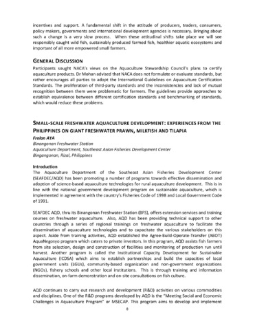
Small-scale freshwater aquaculture development: Experiences from the Philippines on giant freshwater prawn, milkfish and tilapia
(Japan International Cooperation Agency, 2013-12)The Aquaculture Department of the Southeast Asian Fisheries Development Center (SEAFDEC/AQD) has been promoting a number of programs towards effective dissemination and adoption of science-based aquaculture technologies ... -
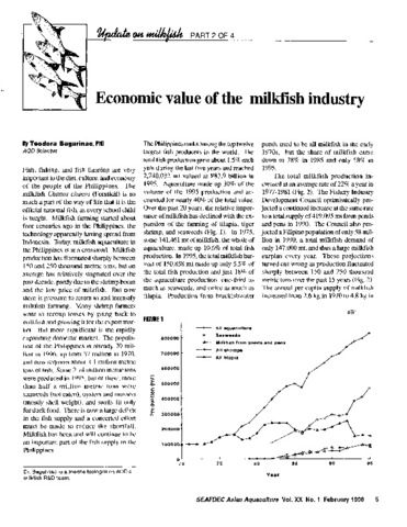
Economic value of the milkfish industry
(Aquaculture Department, Southeast Asian Fisheries Development Center, 1998)A brief description is given of the milkfish (Chanos chanos) farming industry in the Philippines. Over the past 20 years, the relative importance of milkfish has declined with the expansion of tilapia, tiger shrimp and ... -
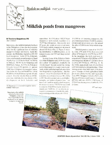
Milkfish ponds from mangroves
(Aquaculture Department, Southeast Asian Fisheries Development Center, 1998)